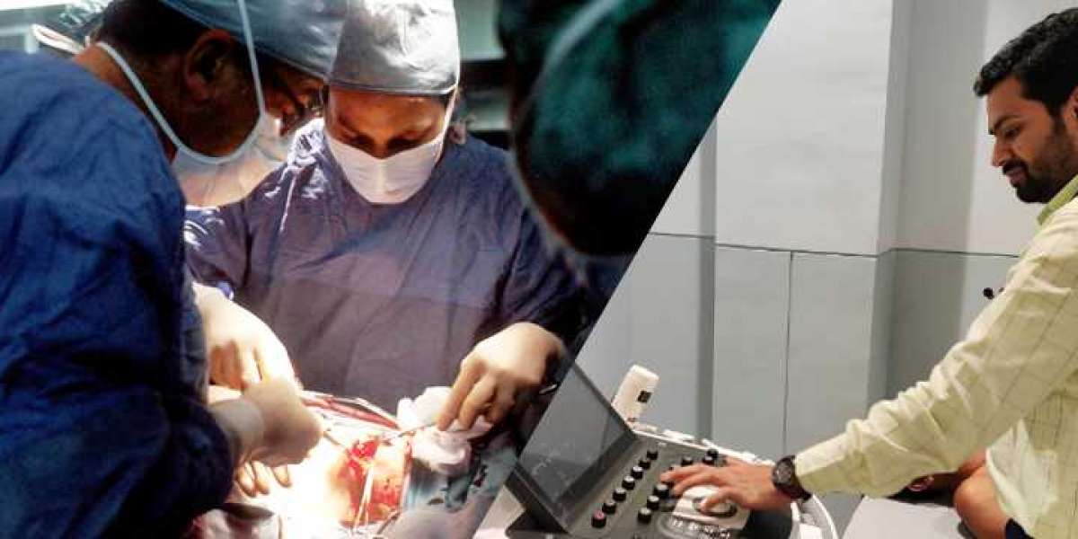
When it comes to heart health, few tools are as valuable as 2D echocardiography. This innovative imaging technique allows us to view the heart in real-time and is instrumental in diagnosing and managing a variety of cardiovascular conditions. At our hospital, we’re proud to be recognized as the best cardiologist in Kawardha and to offer advanced 2D echocardiography services.
What is 2D Echocardiography?
2D echocardiography is an ultrasound-based imaging technique that provides real-time, two-dimensional images of the heart. By sending high-frequency sound waves into the chest, the echocardiogram creates a visual map of the heart's chambers, valves, and blood flow. This allows cardiologists to assess the heart’s structure and function without any invasive procedures.
Why 2D Echocardiography Matters
2D echocardiography offers numerous advantages:
- Non-Invasive: No need for incisions or injections.
- Real-Time Imaging: Provides live views of heart movement and function.
- Detailed Visualization: Offers clear images of the heart's components.
- Safe and Effective: Uses sound waves instead of radiation, making it safe for repeated use.
2D Echocardiography for Adults
Key Uses in Adult Heart Health
For adults, 2D echocardiography is often used to:
- Diagnose Heart Conditions: Identify issues such as valve disorders, heart failure, and cardiomyopathy.
- Monitor Existing Conditions: Keep track of conditions like congenital heart defects that may persist into adulthood.
- Guide Treatment Decisions: Provide detailed images that help in planning surgeries or other interventions.
What to Expect During the Procedure
Here’s a quick rundown of what you can expect during a 2D echocardiogram:
- Preparation: You’ll be asked to change into a hospital gown and lie down on an examination table.
- Application of Gel: A special gel will be applied to your chest to ensure the transducer moves smoothly.
- Imaging: The cardiologist will move the transducer over your chest to capture various angles of your heart.
- Review: The images are analyzed to evaluate your heart's health and function.
After the Procedure
Once the procedure is complete, you can resume your normal activities. Your cardiologist will review the results and discuss any necessary follow-up or treatment options.
2D Echocardiography for Children
Tailored Heart Care for Kids
In children, 2D echocardiography is crucial for:
- Detecting Congenital Heart Defects: Identifying structural issues present from birth.
- Evaluating Heart Function: Checking for any acquired conditions or abnormalities.
- Planning Interventions: Providing detailed images to assist in planning surgical procedures if needed.
How the Procedure Works for Children
For children, the procedure is similar but with extra care for comfort:
- Preparation: Children may need to be calm, so distractions or mild sedation might be used.
- Gel and Imaging: Gel is applied, and the transducer is moved over the chest to obtain images.
- Comfort: Extra steps are taken to ensure the process is as quick and comfortable as possible.
Post-Procedure
After the scan, children usually feel fine and can return to their normal activities. The results will be reviewed by the cardiologist, who will discuss any findings and next steps with the parents.
2D Echocardiography for Fetuses
Importance of Fetal Echocardiography
Fetal echocardiography is a specialized form of 2D echocardiography used during pregnancy to assess the unborn baby's heart. This is essential for:
- Early Detection of Heart Defects: Identifying congenital heart problems before birth.
- Monitoring Development: Tracking the fetus’s heart health throughout pregnancy.
- Planning for Birth: Preparing for any special care needed immediately after delivery.
What to Expect During a Fetal Echocardiogram
Here’s how a fetal echocardiogram typically unfolds:
- Preparation: The mother will lie comfortably, and gel will be applied to her abdomen.
- Imaging: The transducer is gently moved over the abdomen to capture images of the fetus's heart.
- Analysis: Detailed images are reviewed to ensure the baby’s heart is developing normally.
Post-Procedure
Results will be discussed with the parents, including any necessary follow-up or special preparations for delivery.
Why Choose Us?
As the heart specialist in Rajnandgaon, our hospital is dedicated to providing exceptional heart care using the latest technology and techniques. Our team, including the best cardiologists, is committed to offering personalized care tailored to each patient’s needs. Whether you’re an adult, a parent seeking care for your child, or expecting a baby, our advanced echocardiography services are here to ensure the best possible outcomes for your heart health.
Conclusion
2D echocardiography is an invaluable tool in modern cardiology, offering detailed insights into heart health for patients of all ages. From detecting issues in adults to monitoring fetal heart development, this technology helps in diagnosing, managing, and treating various cardiovascular conditions. If you’re seeking top-notch heart care, our team, including the best cardiologist in Kawardha and the heart specialist in Rajnandgaon, is here to provide you with expert care and support.
Feel free to reach out to us for more information or to schedule an appointment. We look forward to helping you maintain your heart health with the best care possible.


